Anatomy120
Monday, June 17, 2013
Thursday, May 26, 2011
Sunday, May 01, 2011
slides: Respiratory System

Wall of Trachea- lumen (right), mucosa (ciliated pseudostrat columnar ep w goblet cells), submucosa ( white layer, red splotchy (tubules + ducts that are part of the glands), white layer), adventitia (dense ct) with it's hyaline cartilage (NEVER IN WALL OF ESOPHAGUS), in esophagus would be muscularis externa (smooth muscle)

Mucosa of last slide- lumen (right), ciliated pseudostrat columnar ep with goblet cells, basement membrane that they are sitting on (whitish stain)

Mucosa of last slide- lumen (right), ciliated pseudostrat columnar ep with goblet cells, basement membrane that they are sitting on (whitish stain)
Lung Tissue- alveolar sacs, alveolar ducts, arteriole (bottom leftish center) with smooth muscle + blood cells in it
Alveoli individ to show caps (hard to find)
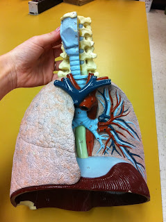
Human lung- alveoli (right side), upper tube blood vessel, layers of smooth muscle + simp squam ep around it, lower tube is bronchial (lined with respiratory epith: cuboidal or columnar)
Pig Dissection
pulling intestine- attached by thin membrane Mesentery, attached to back wall of pig. how intestines are supported in abdominal cavity.
under liver, stomach, membrane connects the two- lesser omentum
slides: Digestive System
Esophagus- strat sq epith (closest to lumen), submucosa (CT) doesn't have all the glands like the trachea, smooth muscle
Esophagus- know it is not trachea bc it is not ciliated pseudo strat columnar ep

Esophagus- strat sq epith, connective tissue underneath, next layer is smooth muscle, NOT cartilage (like in trachea)

Esophagus- strat sq epith, connective tissue underneath, next layer is smooth muscle, NOT cartilage (like in trachea)
Ileum- (small intest), mucosa with villi (upper layer) simp columnar ep, CT inbtwn filling up meat inbtwn villi is lamina propria, muscularis mucousa (tricky to see) is under as a straightish line really thin layer of smooth muscle, submucosa is white and lightly tinted layer thick, muscularis externa is bottom layer but it has been broken
Another at higher mag- serous memb bottom
slides: Digestive System
Intestine wall- he rotated it twice so that yellow hairs are in bottom right corner, top= surface (crypts) mucosa, epithelium is going up and down, columnar cells line the nook near crypts, lamina propria under mucosa, then submucosa with blood vessels and stuff, pink is muscularis externa circular layer, yellow is longitudinal layer, cells inbtwn the 2 layers are neurons of myentary plexus of averbach,
Duodenum- mucosa is upper layer with fingers, second layer is submucosa with glands CT, muscularis externa first layer in it is circular layer then longitudinal layer
Salivary Glands (exocrine)- white big circle things are ducts (simp or strat cuboidal, secretory cells (dark= serous cells, lighter= mucous cells)
Closeup- stratified cuboidal around lumen looking things, mucus cells are more mucusy looking inside, serous demilune (middle of slide), vein is the red thing
transition btwn Esoph + Stomach- lumen, mucosa- strat squam and then simple columnar, submucosa, muscularis
Saturday, April 23, 2011
Thursday, April 21, 2011
models: Lymphatic System
Lymph node model: Axillary, inguinal, + cervical lymph node areas
slides: Reproductive System
ovary- not a human ovary bc there is more than one maturing follicle, mature follicle (right), egg (in middle of follicle)
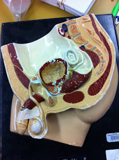
testes- seminiferous tubules, CT inbtwn is where the intersticial tubules cells are
clearer testes- seminiferous tubules, inside the lumen of the tubules are developing sperm, interstitial cell (inbtwn the tubules hard to see)
close up detail of an interstitial tubule- walls of tubule, lumen (sperm production)
slides: Endocrine System
Pituitary gland- neurohypophysis (left; axons) posterior, adrenohypophysis (right dark purple; cells) anterior

Pituitary Gland- top= Anterior Part ("Adrenohypophysis"), bottom= Posterior Part ("Neurohypophysis")

Pituitary Gland- top= Anterior Part ("Adrenohypophysis"), bottom= Posterior Part ("Neurohypophysis")
Thyroid gland w/ follicles (filled with colloid)- T-thyrocytes encircle follicle (make thyroxine), C-thyrocytes (make calcitonin) inbtwn follicles, blood vessel (big upper left), capillary (right)
*only gland that stores products outside
Pancreatic tissue- acinar cells (majority of cells), pancreatic islet of langerhan (upper middle light stained), artereol (right red stained) with blood cells,
duct in pancreatic tissue- simple cuboidal epith lining inside walls Islets of langerhan, acinar cells
*both exocrine (acinar cells) and endocrine (islets of langerhans) "pockets"
pancreas w/ duct
classic duct
models: Endocrine System
Endocrine Baby: upper green= thyroid, red=thymus
green= small intestine, red= pancreas, yellow= adrenal glands
yellow= adrenal glands
Subscribe to:
Comments (Atom)


